Low battery
Battery level is below 20%. Connect charger soon.
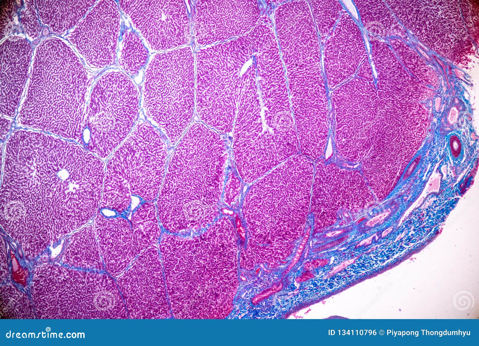
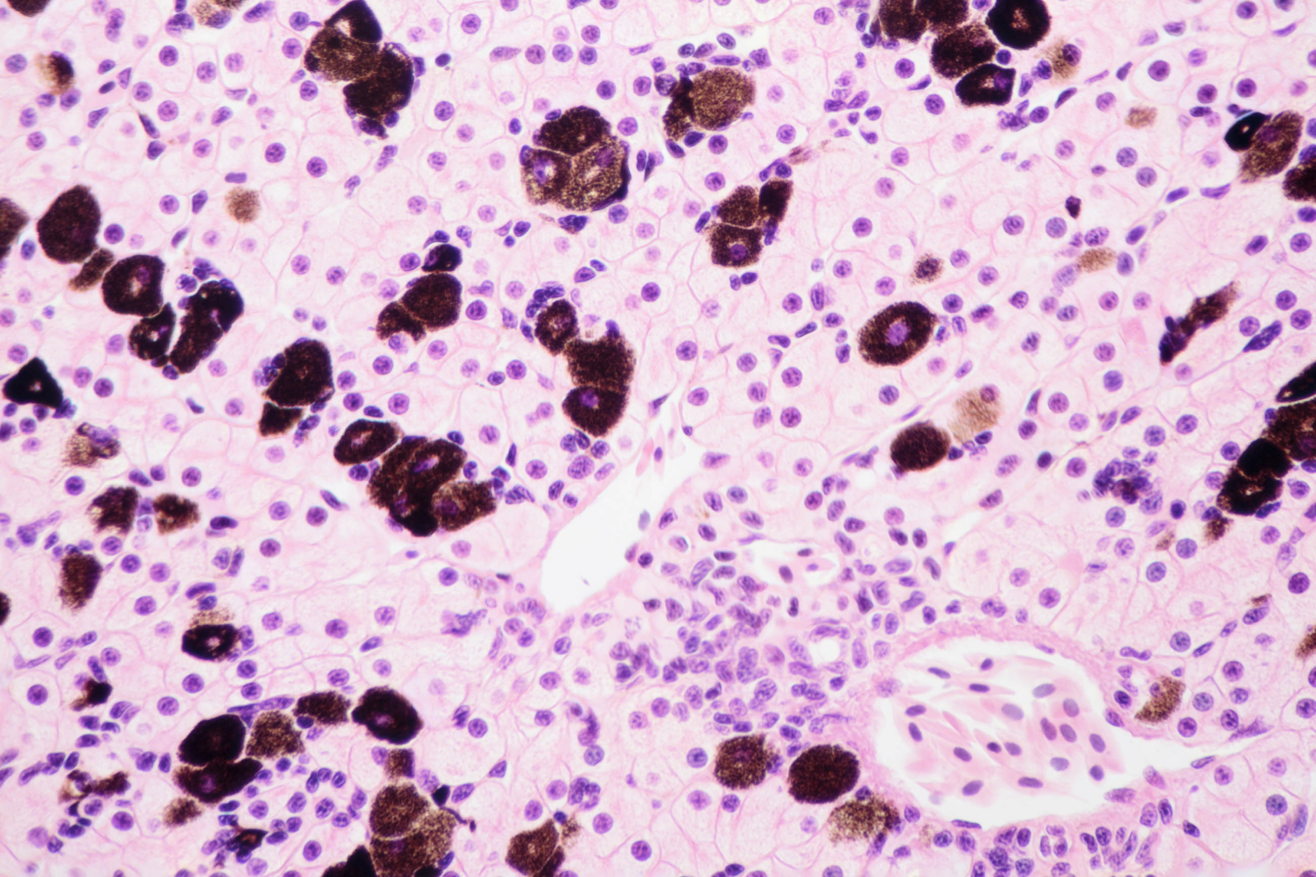
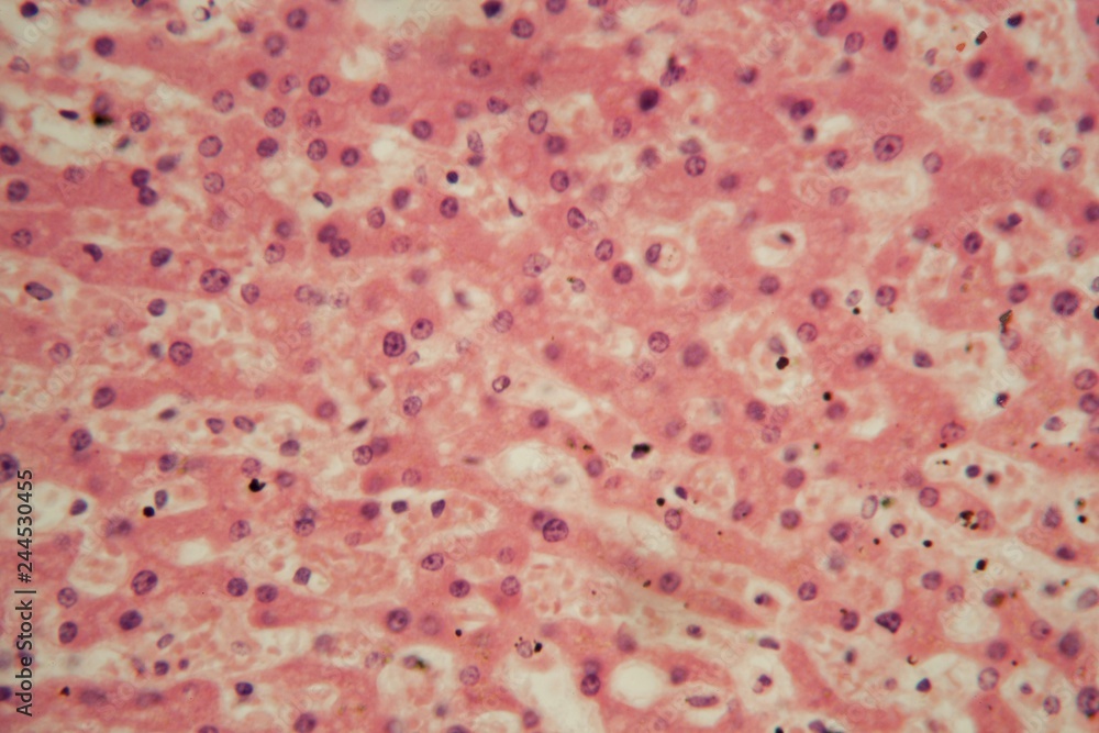
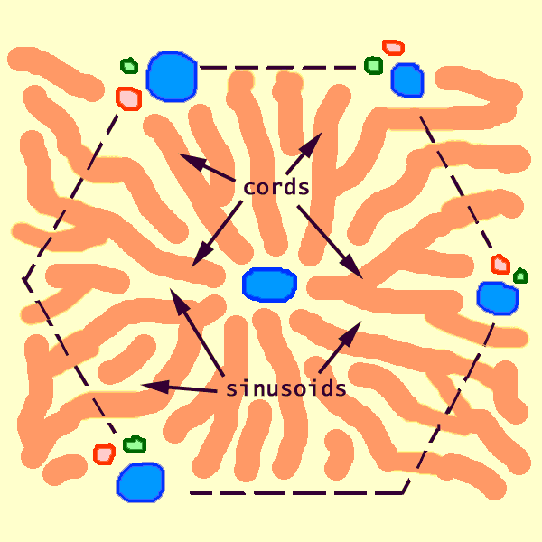

But every cell also has unique features to do a specialized. This article will show you histology slides from the following different organs. The onion peel cell experiment is very popular for observing a plant cell structure. The liver is a large organ in the abdomen that performs many important bodily functions, including blood filtering. Do they look like cell diagrams you’ve seen? · they can view very small specimens and distinguish their structural differences, for example, the view of animal and plant cells viewing microscopic bacterial cells. They highlight a set of overlapping features that all cells need to live. This post explains the theory, requirements, procedure, observation, result and precautions of the onion peel cell experiment. · histology slides guide histology is a visual and colorful science that is studied with the help of a light microscope. Home electron microscopy chapter 15 - liver and gallbladder micrograph information index search contact us … View the leaf under low, medium, and high power objectives, and then draw the cells in figure 2. 2, along with any organelles you can see. · the histology of the pancreas can be studied by staining sections of pancreatic tissue and viewing them under a microscope these stained samples can then be examined for drawing and labelling to identify the exocrine and endocrine tissues of the pancreas If you have difficulty printing out this page, try the pdf version: Electron beams accelerated. Real cell gallery the images in this gallery show real cells under the microscope. · the liver has a lot of vital tasks including ridding the body of toxins. There are different types of microscopes like light microscope, dark-field microscope, phase contrast microscope, electron microscope, fluorescent microscope, etc. It is also considered a … The problem of the limited resolution of the traditional light microscope was overcome in the 1930s, by the development of electron microscopes, which use beams of electrons instead of light. Be sure to label the chloroplasts, the cell membrane, and the cell wall. Electron beams have a shorter wavelength than that of visible light, and the wavelength of a beam of electrons decreases as the velocity (speed) at which they travel increases. Most people do not have symptoms until a late stage. Find out about the different types of liver disease and their causes. Find out the symptoms of liver disease, when to get medical help and what … Most cell diagrams, whether in your textbook or online, are generic. · you are expected to be able to identify free ribosomes, rough endoplasmic … I hope you have a piece of good knowledge of permanent slide preparation, properties of different staining, and handling of the microscope. If you could be at risk, dont wait for symptoms. The diagnosis of liver disease is made by liver function tests, groups of blood tests, that can readily show the extent of liver damage. Learn about problems that can affect the liver and how to … These are the prerequisites to study any histology slide. Liver disease develops slowly.
